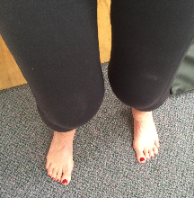
The effectiveness of healing exercise is based on correcting and conditioning your mechanical/postural faults. To do this, you must first identify what these faults are. A basic self-analysis of your body is presented here to allow you to discover what faults you may have that are at the root of your pain and injury. More detailed analysis of any specific body region is provided in any of the Pain Free & Fit TM Healing Exercise Series program books. Once you have identified where your mechanical and postural problems are, you can then choose the appropriate corrective exercises to assist your body's healing process.
The self-analysis consists of two parts- posture/alignment and body mechanics. Take your time with this analysis, as the information you gain here will be used in every pain relieving and healing exercise that is to come. Write down your findings and burn them into your permanent memory. A checklist and video tutorial is provided at the bottom of this page. You may want to enlist the help of a friend to make your analysis easier. Two sets of eyes usually see more detail than one, and a friend can help you confirm or discard a questionable finding.
PART ONE
Analysis of Posture and Alignment
Stand in front of a full length mirror for this analysis. Assume your most natural posture.
1. Foot Posture. Ideally, your feet should be pointing straight ahead, with your body weight being equally distributed on the four corners of each foot (the big and small ball of your forefoot, and the inner and outer corners of your heel). If your feet lean more on the inner edges or inner heel (foot pronation), your Achilles tendon at the back of your heel will bow inwards. Conversely, if you lean with more weight on the outer aspect of your feet (foot supination), your Achilles tendon will bow outwards. If you have trouble checking this, ask a friend to view you from behind. If your heel leans in or out, your Achilles will not be vertically straight and easily seen from behind to bow in or out. If you do not see any problem standing, check your feet again as you sit back (squat) with your hands over-head. This maneuver will often bring out a "hidden" foot posture problem. Be certain on this aspect of the analysis- it is huge for correcting the foundation of your entire body structure.
Look at the top of your feet, just behind your toes. Normally, this region should have a slight raised contour, indicating a normal transverse arch underneath your forefoot. This arch runs from one side of your forefoot to the other, and is located just behind the balls of your feet. If this region looks flat, and especially if it looks concave (similar to a valley or hollow), it indicates decreased transverse foot arch posture.

Pronation Supination
Decreased Transverse Foot Arch
2. Hip-Leg Rotation Posture. In normal standing posture, your knee caps and feet should ideally point straight ahead or very slightly out to the sides. Your right and left legs should be symmetrical. If one or both knee caps and feet point outwards, you have an externally rotated hip posture. If they point inwards, you have an internally rotated hip posture. External hip rotation is much more common than internal hip rotation, and is one of the most important postural faults that can affect many different joints and musculo-skeletal pains throughout the body. It is often the mechanical root cause of many knee, spine and shoulder problems. Healing exercise programs for the entire body have been built around this one foundational fault. Be absolutely certain with this part of your analysis, as it will be the key to many pain relieving exercises. Note: if both your knee cap and the "bony bump" (tibial tuberosity) located 1 inch below your knee cap point straight forward, but your foot turns out or in, you do not have a hip rotation, but rather a tibial torsion of your lower leg, or a foot deformity. In the standing picture below, the model's right hip (viewers left side) is externally rotated.
Right Hip External Rotation Confirmed Right Hip Ext. Rot.
External hip rotation can be slight, but nevertheless very important. If you are not sure about this part of the analysis, close your eyes and march in place for 20 steps. Then open your eyes and look at the alignment of your knees and feet. If they are not pointing straight ahead, you have hip rotation. A confirming test for hip external rotation posture is performed by lying face down and bending your knees as demonstrated in the picture above on the right. While you keep your knees together, allow your ankles to fall as far out to the sides as is comfortably possible. The side which your ankle does not fall as far outward to is the side of external hip rotation. This is on the model's right leg in the picture above. The only exception to this confirming test is if you have an orthopedic condition known as "femoral anteversion or retroversion", which would not be considered as a hip rotation posture discussed here. These are unusual conditions which can be confirmed by an orthopedic physician with a simple in-office test. DO NOT perform hip rotation corrective exercises if you have femoral anteversion or retroversion.
3. Front View of Posture Alignment. Return to your standing analysis, viewing the front of your body in a mirror. Your hips, ribcage, shoulders, and eyes should be level from right to left sides. If one side is higher or lower, they are not level, and you have a side tilting posture of your head, shoulders, or hips. Your hips, ribcage and head should also be stacked vertically on one another in alignment. This should resemble three equal sized boxes all stacked in a column. Note if one of these areas is positioned sideways and not in vertical alignment with the other regions (as if one of the boxes has moved sideways in relation to the others). This misalignment is known as lateral translation posture. There should be no shoulder, hip or foot that is positioned more forward than the other side. The foot of an externally rotated hip will often be positioned more forward than the opposite foot while standing, or on top while crossing the legs. Your face, torso, and pelvis should be positioned straight ahead, and not turned (twisted or rotated posture) to one side. Check for even subtle amounts of rotation in your face (head), shoulders, chest and hips (pelvis). It helps to place a finger on these areas to highlight any rotation (turning to one side).

4. Side View of Posture Alignment. Now turn sideways in front of the mirror and view your standing posture from the side. The center of your ear, shoulder, hip and knee should normally all be vertically aligned when viewing your body from the side. That same line should pass just in front of your ankle while standing. If the center of your ear is more forward than the center of your shoulder, you have forward head posture, which increases neck, back and shoulder stress. Forward hips (sway back posture) are associated with weak "core" and buttock muscles, which cause abnormal lower back, hip and knee stress.
Now check your pelvic alignment. Normally, your pubic bone should be level with the tip of your tailbone. Place one finger on your pubic bone, and the other on the very end tip of your tail bone. Compare the height of your fingers while viewing yourself standing sideways in a full length mirror. They should be level in normal posture. Is your tailbone higher than your pubic bone (anterior pelvic tilt posture)? This will increase the arch (or curve) of your lower back, and can stress your lower back, hips, knees and neck. Is your pubic bone higher than your tailbone (posterior pelvic tilt posture)? This causes a decrease in your lower back arch and can stress the same areas just mentioned.
Sway Back Posture
5. Shoulder & Upper Arm Posture. Alignment of your shoulders consists of both your shoulder blades and your upper arms. Begin with examining your shoulder blade alignment. The front of your shoulders should be flat in contour. If your front shoulder contour is hollowed, concave, or "scooped out", you have rounded shoulder posture. One shoulder blade should not rest higher on your ribcage than the other. If it does, you have elevated shoulder posture. This may not be apparent until you lift your arms. Normally, your shoulder blade should rotate, but not elevate as you raise your arm sideways, until your arm is reaching maximum heights. A friend can feel your shoulder blades from behind to help you with this analysis. Normally, the top of your shoulder blade should be level. If the outer aspect of the top of your shoulder blade is lower than the inner aspect, you have downward rotated shoulder posture. This often causes your shoulder to appear lower than the opposite side. If the outer aspect of the top of your shoulder blade is higher than the inner aspect, you have abducted shoulder posture. This is usually associated with the arm on the same side being held farther away from your torso than the opposite side. If the lower tip of your shoulder blade is not held flat against your back, but "pops out", you have a tipped shoulder posture. Finally, if the entire inner edge of your shoulder blade "pops out" away from your back, you have a winged shoulder blade posture.
To analyze your upper arm posture, feel the contour of the front and side of your shoulder. If you feel that the front bulges forward, you likely have an anterior upper arm posture. If the side contour feels higher than the opposite side, you likely have a superior upper arm posture. Both of these postures consist of an abnormal placement of your upper arm in your shoulder socket, and while alignment with your arm hanging at your side can be informative, feeling this area during motion is more confirming of these mechanical flaws. In normal posture, the palms of your hands should point in towards each other, not backwards. The points of your elbows should point backwards and not sideways. Palms that point backwards (and elbows that point outwards) can indicate rounded shoulder posture, internal rotation posture of the upper arm, or tight latissimus dorsi muscles. All of these can stress the shoulder, neck and lower back regions. To distinguish between these, hold your shoulder blade so that the front of your shoulder has a flat contour. This corrects any rounded shoulder posture. If the palm of your hand still faces backwards, you have an internal rotation posture of your upper arm. In the picture above, the model has slight rounded shoulder posture, and major internal rotation posture of his upper arm. Test for latissimus dorsi tightness by laying on your back with your legs straight. If you cannot touch the floor by reaching overhead without arching your lower back, your latissimus muscles are definitely tight.
PART TWO
Analysis of Body Mechanics
Now that you have a good picture of your posture, it is time to see how these findings relate to your body mechanics. Good mechanics are demonstrated by proper joint stability and muscular coordination. These ensure proper use of your musculo-skeletal system, which decreases stress and injury potential. By identifying where your mechanical problems exist (and then learning to correct them), you can greatly decrease the mechanical stresses that are causing your pain.
Body mechanics are best analyzed by using both a global test to determine the weak links of your full, multi-joint body system as a whole, and with several local tests to determine the stability of specific body regions. As you perform these tests, be sure to record your findings. They will be used together with your posture findings to construct your body-specific corrective healing exercises.
Add Text Here...
Global Analysis
The Over-Head Lunge Maneuver. Stand in front of a full length mirror, or enlist a friend to watch your mechanics for this test. You will use the over-head lunge maneuver as the first mechanical test for your analysis. This maneuver puts your entire body in an unstable position and highlights where your alignment and stability problems exist. It is an analysis of your global (full body) posture and stability. You will be looking for any unbalanced movement or alignment tendencies. Begin by reaching over-head and step forward with one leg. Allow your rear heel to raise off the floor. Allow both of your knees to bend as far as possible, taking note of any unbalanced movement or alignment. If you begin to feel unbalanced, note the first movement that your body makes. Do not record any secondary movements as you try to re-balance yourself. You are only interested in your body's first unbalanced movement tendencies. As you bend your knees and descend into the over-head lunge position, check all of the following points to begin a list of your mechanical faults:
1-Did your lead hip move upwards (pelvis swing up towards the ribs on the lead side)? Normally, the hips should remain level. If your lead hip moved upwards, you have identified the hip hiking mechanical fault on that side.
2-Did your pubic bone turn away from your lead leg? Normally, it should still be facing straight forward. If it turned away, you have the mechanical fault of pelvic rotation to the side it is turning towards.
3-Did your lower back arch increase? Normally, the curve in your lower back should remain motionless during this maneuver. You may use one of your hands to monitor this as you perform the lunge. If the arch increases, you have the mechanical fault of lower back hyperlordosis. This is usually associated with your tailbone moving higher than your pubic bone (anterior pelvis). Use your fingers on your tail bone and pubic bone to monitor this.
4-Did your torso or shoulders lean or twist to one side? Normally, they should not lean (tilt) or twist. If they lean/tilt to the right, your mechanical fault is right torso side tilting. If they lean to the left, your tendency is left torso side tilting. If your chest twists to the right, you have right torso rotation, and if it turns to the left, you have left torso rotation.
5-Did your head move forward so that your ears are in front of your shoulders (forward head posture tendency)? Normally, your ear should remain vertically in line with your shoulder during the lunge.
6- Did your head or neck tilt sideways (head-neck side tilt posture), or did your face turn to one side (head-neck rotation)? Normally, your head and neck should remain straight.
7- Did your shoulders round (rounded shoulder posture) or move up (elevated shoulder posture) towards your ears?
8-Did your front heel/ foot tilt inwards (pronation) or outwards (supination)? Normally, the heel should stay balanced with equal weight on the inner and outer edges.
9-Did your front knee move inwards (knee valgus mechanical fault), or outwards (knee varus mechanical fault)? Normally, the front knee should remain centered over the ankle as it bends and should not move in or out.
10- Do your knee caps face the same direction as your toes do? Check this on both the front and rear legs. Normally they should. If they do not, you have a knee rotation mechanical flaw.
11- Did your front knee move forward past your front toes? Normally, it should not move forward past the tips of your toes. If it does, you have a forward knee mechanical fault on your lead leg side.
12- Are your wrists more forward than your shoulders? Normally, you should be able to hold them directly over your shoulders with your elbows bent to 90 degrees. If you cannot rotate them into this position in the lunge, you have identified internal rotation posture of your shoulder(s).
Make note of any of the above mechanical faults as you descend into the lunge, and also as you hold the bottom position of the lunge for 10-15 seconds. Sometimes a mechanical fault will appear during descent, and sometimes it will show itself while holding the bottom position. Note any of the above findings and then repeat the test with your other leg stepping forward. Many of your findings may match your standing posture analysis findings, while others may be new discoveries. If you cannot find any of the above alignment tendencies, try the same test again with your eyes closed (enlist a friend to help with this analysis). Closing your eyes will help you discover slight mechanical faults that may not be apparent during a typical standing posture or lunge analysis.
Local Analysis
While the over-head lunge tests full body global alignment and stability function, the following local analysis tests will help you to identify region-specific mechanical stability and coordination faults. To maximize the effects of any Pain Free and Fit TM Healing Exercise Program, you will want to correct both global and local mechanical problems. This is essential before beginning any conditioning (strength, endurance, flexibility or balance) training program. Without identifying and correcting your mechanical faults first, you will only strengthen dysfunction. By correcting mechanics and knowing what to watch out for in terms of mechanical problems, you can get the full pain-relieving and healing-stimulus effects of the routine.
1. Drawing-In Abdomen and Glute Bridge Test. This first local mechanical test is done lying face up with your knees and hips bent. Your feet are flat on the floor. Begin the first part of the test by placing your fingertips two inches to the sides and one inch below your belly button. Do not press in deeply as you want the touch to remain soft to sense what you will do next. Gently pull (suck-in) your lower abdomen toward your spine. Feel your lower abdomen move down and away from your fingers. Do both sides move away equally? Does one side move less? Does one or both sides bulge out toward your fingers?
The deepest of your abdominal muscles (known as your transverse abdominus) will draw your lower abdomen away from your fingers equally on both sides if you have proper mechanics. If you feel one side drawing in less, or bulging outward, it indicates a compensatory internal oblique muscle contraction. Make note of which side this mechanical fault occurs on, if it exists. The abdominal oblique muscles are more superficial to the transverse abdominus and should not contract with this draw in maneuver under normal circumstances. Internal oblique contraction causes a bulge outwards instead of a hollowing inwards with this test. This is a sign of improper mechanics, and demonstrates incorrect abdominal muscle stabilization of your lower back and "core". Record your findings and proceed to the second part of this test.
From the same position, lift your hips off the floor as high as is comfortable (Glute Bridge Maneuver). Hold this top position and compare the "tightness" or "hardness" of your lower back erector spinae muscles to your buttock muscles. Your lower back erector spinae muscles are located 1-2 inches away from the midline of your spine on both sides. Use your fingers to judge the comparative "hardness" or "softness" of each muscle.
The buttock muscles should normally feel slightly harder than the lower back muscles. If you feel more hardness or tension in your lower back, it indicates compensatory "over working" lower back erector spinae muscles for weak buttock muscles. This is a very common mechanical fault, and is often responsible for lower back and hip pain. The buttocks are used for every step you take, every stair you climb, and every time you get up from a stooped or seated position. When the buttocks (gluteus max muscles) are weak, the lower back will compensate and over-work (strain) in an attempt to help the weak buttocks do their mechanical job. The erector spinae are smaller muscles which were never designed to work that hard, and as a result, place excessive stress on spinal joints and discs. Weak buttocks cannot support proper hip function, and cause a compensatory over-stress to other hip muscles and soft tissue structures.
As you hold the top position, look at your hip alignment. Check to see that your hips are not rotated (one hip higher than the other), or tilted (one hip closer to the bottom of your ribs "hip hiking" than the other) in this position. Previous posture findings often are evident during this test. Record your findings.
2. Hip Flexion Test. This local test of hip joint mechanics is also done lying face up, but with your legs straight out on the floor. It also has two parts. The first is done by feeling the hardest and most prominent aspect on the side of your hip. This is your greater trochanter bone. It is roughly the size of a golf ball. You can't miss it, as it is the only hard point on the side of your hip.
Hip Flexion Test
Feel the front of this bone with your index finger, and the back of it with your middle finger of the same hand. Place your opposite hand under your lower back. As you bend your knees (try one at a time and then both simultaneously), you will be checking for two mechanical mistakes. The first will be an arching of your lower back. This will be apparent if your lower back moves away from, instead of into, the hand under your lower back. With normal mechanics, the low back should either not move at all, or flatten your hand. If your spine moves away from your hand, it indicates poor abdominal control of your lower back during hip flexion (bending your hip and knee forward). This mechanical flaw is called low back hyperlordosis tendency, and may confirm an earlier finding in the over-head lunge test.
The second mechanical mistake with this test will be if your greater trochanter moves forward into your index finger. It should normally tense slightly backwards into your middle finger (staying centered in the hip joint). Movement forward (anterior hip malposition posture) indicates an imbalance in hip flexor-extensor muscle activity which contributes to tight hips, weak buttock muscles, and an overarching of the lower back curve (hyperlordosis). It is also commonly found on the same side as an externally rotated hip posture. Any one (or all) of these will stress various lower back, hip and knee structures, contributing to pain in these areas.
3. Multifidus Test. The next test analyzes the mechanics of the multifidus muscle. The multifidi are key stabilizing muscles that attach to your lower back vertebra, protecting your lower back from pain. They are an essential component of your "core" muscles. Begin standing and place your thumbs against the sides of the "knobby bumps" of your spine located at the midline of your lower back. The exact location you want to feel is the lower/outer tips of each of these vertebral bones. You have five of these in your lower back, so feel each one and try to determine if you feel any difference from right to left sides, or from one vertebra to the next. You are feeling for a "hollow" or "depression" at the lower/outer tip of each vertebrae which indicates a weak (undeveloped) multifidus muscle. Now feel the same areas as you draw your lower abdomen inwards to your spine. Do not move your spine during this test.
Multifidus Test
4. Knee Cap Tracking Test. Lying face up, or sitting on the floor with your legs out straight, place your two index fingers on the top of your knee cap. One finger will be contacting the upper/inner border of the knee cap, while the other contacts the upper/outer border of the knee cap. As you slowly straighten your knee, determine if you feel the same tension at both of your fingers. Normally, your quadriceps (front thigh) muscle tenses as you straighten your knee, causing the knee cap will move up (towards your hip) into both of your fingers with equal pressure.

In lateral tracking patella disorder (weak VMO), you will feel more tension on your outside finger as your knee cap moves outward while your quadriceps tense and the knee straightens. If you feel more tension with your outside finger, or feel the knee cap push against your outside finger more than your inside finger, you have a lateral tracking knee cap (patella). This means that the quadriceps muscle located on the inner edge of your knee cap (the VMO muscle) is weaker than the quadriceps muscle on the outer aspect of your knee cap. This imbalance causes your knee cap to shift sideways towards the outside of your knee. This is a common cause of front knee pain. You may also feel the inner edge of the knee cap move forward with this test, which would indicate a tilted patella dysfunction. Both tilted and lateral tracking patella problems are the result of weak VMO muscles, externally rotated hips, anterior pelvic tilts, and pronated feet. These commonly occur together, and require healing exercises which address all of these problems for maximal pain relief and correct mechanics. If you feel equal tension and movement between your fingers, you do not have a tracking problem. In some instances, the inside finger will have more tension, which indicates a medial (inside) tracking patella. Record either of these mechanical faults if they are found.
5. Knee Rotation Test. As the knee bends, the knee cap should ideally point in the same direction as the foot points towards. With the mechanical error of knee rotation, the knee cap faces a different direction than the foot while the knee bends. This is analyzed using the pivot squat test. The pivot squat test is performed by raising your heels and turning both feet to one side by pivoting on the balls of your feet, and then bending both knees. Hold this bent-knee position as you check for the alignment between each foot and knee cap. If the foot and knee cap face different directions, knee rotation exists. If your foot faces outwards compared to your knee cap, you have external knee rotation. If it faces more inwards, you have internal knee rotation. Perform this test to both sides. If the knees are positioned inwards compared to the rest of the upper and lower leg, knock knee (valgus) knee posture exists. If they are positioned outwards, bow legged (varus) posture exists.

External Knee Rotation and Knock Knees (valgus)
6. Breathing Test. Place one hand on your chest, and the other on your upper abdomen (above your belly-button). Under normal breathing conditions (including light exercise), your upper abdomen and lower ribs should move as you breathe. Your chest should not move, or only move minimally. Your diaphragm muscle separates your lungs and heart cavity from your abdomen. It is an essential "core" muscle. Breathing mechanics are normal if your upper abdomen and lower ribs move, while your chest remains motionless during minimal exertion. If you feel your chest move, or if it moves more than your abdomen during heavy exercise, you have the mechanical fault of chest breathing. This can cause an overstress to your neck region as neck and chest muscles are over-compensating for decreased diaphragm movement. This often causes neck and shoulder pain. The only time the chest should move considerably is with moderate to heavy physical exertion in order to increase lung volume and breathing capacity.
7. Neck Stability Test. Lying on your back with your hips and knees bent, and your feet on the floor, look at your knees. If your head barely came off the floor, but rotated to allow your eyes to look at your knees, you have normal neck stability mechanics. If your head jutted forward and came up off the floor to look at your knees, you have weak deep neck flexor muscles, which is a mechanical flaw in neck stability.
Normal Neck Stability Weak Deep Neck Flexors
8. Shoulder/Upper Arm Stability. Shoulder stability relies on both shoulder blade and upper arm position. The shoulder blade has already been analyzed. This test determines if the upper arm is centered properly in the shallow shoulder socket with motion. Feel the front of one of your shoulders as you raise that arm forward. Normally, the upper arm should move slightly backwards or stay centered in the shoulder socket during this motion. If you feel the upper arm moving forward into your hand, an anterior upper arm malposition is the mechanical fault. Check both shoulders. If the "crook" or "bend" of your elbow turns slightly inwards (towards your chest) as you raise the arm, you have discovered an internal rotation upper arm malposition, as it should normally have no rotation at all with raising to about shoulder level, or turn outwards when raising above shoulder level. Subtle mechanical flaws in upper arm position can be better appreciated by feeling both shoulders at the same time and comparing them as the arms are raised. A friend can help you by feeling both of your shoulders as you raise your arms simultaneously. This will often highlight an anterior or internal rotation movement which may have not been as noticeable with single arm testing.


Anterior Upper Arm Testing Superior Upper Arm Testing
The upper arm should also be tested for superior (upward) malposition. For this part of the analysis, feel the upper aspect of the side of your shoulder. You will want to feel the side of your shoulder just off the bony point on the top of your shoulder. As you raise your arm sideways, feel for an abnormal upwards movement of the upper arm. With normal mechanics, the upper arm should remain centered in the socket, or move slightly downwards as the arm is raised sideways. If the upper arm shifts upwards, a superior (upward) upper arm malposition mechanical error has been found.
By combing these basic global and local analysis tests, you now have found where your major mechanical faults or "weak links" are located. Combining this knowledge with your previous alignment/posture findings found above, or in the Quick Check Video at the top of this page, gives you a basic "whole body" picture of the structural and functional distortions that are most likely contributing to your pain. While this will give you a great starting point for your healing exercises, a more detailed analysis per body region is available in any of the Pain Free & Fit TM Self- Help Healing Exercise Series e books. The more detailed and thorough your analysis is, the greater the corrective and healing potential is with your exercises. The checklist below will help you in recording all of your basic findings. This information is essential for creating and performing any Pain Free and Fit TM healing exercise, as conditioning your posture/alignment and mechanical fault corrections is the basis for correctly stimulating your body's healing mechanisms. With this in mind, you are now ready to learn some healing exercises available through any of our products, sample videos of our Online Healing Exercise Workout, or through our you tube channel. Enjoy!
Pain Free & Fit TM / Posturcise ®
Posture and Body Mechanics Self-Analysis Check-List
Copyright 2015, Dr. Michael C Remy, North Shore Health & Fitness Associates, Inc.
All rights reserved.
1. Foot Posture
__Pronated
__Supinated
__Decreased Transverse Arch
2. Hip/Leg Rotation Posture
__ External Rotation
__Internal Rotation
3. Side View Posture
4. Front View Posture
5. Shoulder Posture
__Winging Shoulder Blade
__Elevated Shoulder Blade
__Downward Rotated Shoulder Blade
__ Abducted Shoulder Blade
__Tipped Shoulder Blade
__Anterior Upper Arm
__Internal Rotation Upper Arm
6. Global Mechanics (Over-Head Lunge Test)
__Pelvic Rotation
__Lower Back Hyperlordosis
__Heel Supination
__Torso Side Tilting
__Torso Rotation
__Knee Rotation
__Forward Knee
__Head-Neck Side Tilt
__Head-Neck Rotation
7. Local Mechanics
__Weak Gluteus Max with Erector Spinae Compensation
__ Low Back Hyperlordosis
__ Anterior Hip Malposition
__Weak Multifidus
__ Lateral Tracking Knee Cap
__ Medial Tracking Knee Cap
__ Knee Rotation
__ Chest Breathing
__Weak Deep Neck Flexor
__Anterior Upper Arm Malposition
__Superior Upper Arm Malposition
__Internal Rotation Upper Arm Malposition
Self -Analysis of
Posture & Body Mechanics


